
Environmental hypoxia exerts detrimental effects on the reproductive capabilities of both humans and animals. A fetal hypoxia model was established in which fetal mice were kept in a high-plateau hypoxic setting from embryonic day (E) 0 to 16.5. In our previous research, we found that fetal hypoxia exposure perturbs the methylation of imprinted genes in adult sperm and causes intergenerational placental impairments in male offspring. However, the specific impacts of fetal hypoxia on the female reproductive system, particularly with regard to oocyte maturation, remain poorly understood. Firstly, we found that fetal hypoxia mice exhibited a significant reduction in the average number of pups per litter. We conducted a comprehensive analysis of the transcriptome in oocytes from the hypoxic group and investigated the metabolic alterations within the follicular microenvironment. Fetal hypoxic stress contributed to cleavage and blastocyst rate reduction and induced early apoptosis and DNA damage triggered by mitochondrial dysfunction, oxidative stress aggravation and Sirt3/Sod2 down-regulated. Additionally, administration of nicotinamide mononucleotide (NMN) has been shown to prevent oocytes from mitochondrial dysfunction and developmental impairment by increasing expression of Sirt3/Sod2 and autophagy. The number of pups per litter in fetal hypoxia mice was reduced by 57.7% compared to the control group, while NMN intervention could restore it to 73.1% of the control group. These results indicate that fetal hypoxia exposure exerts multiple potential damages to adult female reproduction, while highlighting the clinical potential of NMN supplementation as a targeted intervention to alleviate such hypoxia-associated female reproductive impairment.
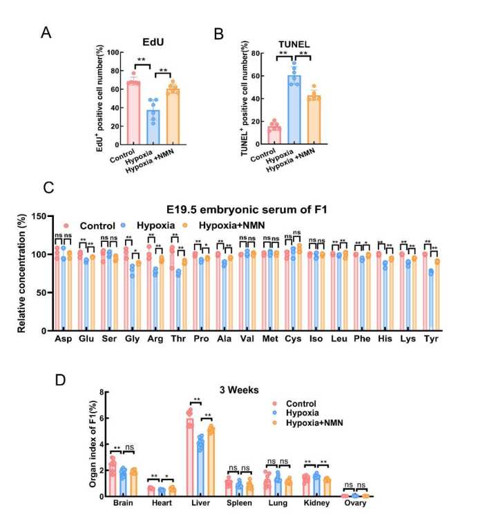
Figure S10 A: EdU+ positive cell numbers in Control, Hypoxia, Hypoxia + NMN ovaries. B:Tunel+ positive cell numbers in Control, Hypoxia, Hypoxia + NMN ovaries. C: Amino acid of F1 E19.5 embryonic serum in Control and Hypoxia. D: Organ index of 3 weeks in F1 Control,Hypoxia, Hypoxia + NMN. Results are presented as mean ± SD. ns: Not significant; *:P < 0.05;**:P < 0.01.
The link below will guide you to the reading:
https://www.zoores.ac.cn/article/doi/10.24272/j.issn.2095-8137.2025.579